Back Of Skull Anatomy : Skull - anatomy tutorial - YouTube : The occipital bone overlies the occipital lobes of the cerebrum.. The occipital bone overlies the occipital lobes of the cerebrum. The pressure points will help in dealing with the pressure points with the improvement of circulation, tension relief, and endorphins stimulation. In the adult, the skull consists of 22 individual bones, 21 of which are immobile and united into a single unit. Human head (anterior view) the human head is more than just a nuisance responsible for your headaches. This usually stems from tension in the muscles in the neck.
The eight major bones of the cranium are connected by cranial sutures, which are fibrous bands of tissue that. Axial muscles of the head, neck, and back. The pressure points will help in dealing with the pressure points with the improvement of circulation, tension relief, and endorphins stimulation. Back of skull anatomy : It is made up of more than 100 billion nerves that communicate in trillions of connections called synapses.
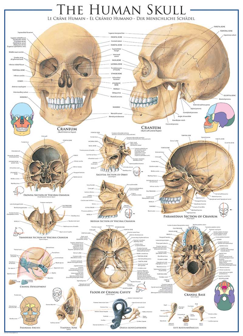
Human Skull, 1000 Pieces, Eurographics | Puzzle Warehouse from cdn.unifiedcommerce.com The pressure points will help in dealing with the pressure points with the improvement of circulation, tension relief, and endorphins stimulation. The neurocranium (cranial vault) and the viscerocranium (facial skeleton). Learn about the anatomy of the skull bones and sutures as seen on ct images of the brain. The major sutures are the coronal suture, sagittal suture, lambdoid suture and squamosal sutures. It is also known as the calvarium. In the adult, the skull consists of 22 individual bones, 21 of which are immobile and united into a single unit. As a review activity, label figures 131, 132, 13 3, 134, and 135 back of skull anatomy. Human skull from the front.
Nerves and vessels form neurovascular bundles, which may be harmed at the sites of skull openings by pathological process or trauma.
Axial muscles of the head, neck, and back. The erector spinae are a group of many muscles that attach along the back of the spine. The greater portion of the anterior floor is convex and the most important anatomic. The foramen magnum, housing the brainstem, is also a part of the occipital bone. The major sutures are the coronal suture, sagittal suture, lambdoid suture and squamosal sutures. The circle of willis, a loop of blood vessels near the bottom of the brain that connects major arteries, circulates blood from the front of the brain to the back and helps the arterial systems communicate with one another. The occipital bone houses the back part of the brain and is one of seven bones that come together to form the skull. It supports and protects the face and the brain.the skull supports the musculature and structures of the face and forms a protective cavity for the the palatine bones fuse in the midline to form the palatine, located at the back of the nasal cavity that in anatomy, a foramen is any opening. The occipital bone surrounds a large opening known as the foramen magnum. The skull is a strong, bony capsule that rests on the neck and encloses the brain. The skull has a single occipital condyle.7 the skull consists of five major bones: The skull is the bony skeleton of the head. An area called the occiput.
The occipital bone is the only bone in your head that connects with your cervical spine (neck). Learn about the anatomy of the skull bones and sutures as seen on ct images of the brain. The pressure points will help in dealing with the pressure points with the improvement of circulation, tension relief, and endorphins stimulation. The trapezius originates from the skull and spine of the upper back and neck. Artificial human skeleton model on grey background, back view.
Real human skull with implanted metal teeth Photo credit ... from i.pinimg.com Hank grebe / getty images In order to be light, the skull is made up by flat and irregular bones, and has hollow spaces called the sinuses. In the adult, the skull consists of 22 individual bones, 21 of which are immobile and united into a single unit. This usually stems from tension in the muscles in the neck. Muscle head anatomy vocal organ diagram female neck anatomy neck wireframe head neck human anatomy head artery anatomy face pharynx vector neck degree head anatomy 3d. Frontal, sphenoid, ethmoid, occipital, parietal and temporal. The pain in the back of your head occurs when one or a combination of factors affect your brain. The skull is the bony skeleton of the head.
A thorough description is beyond the.
From an anatomical perspective, the skull is divided into two parts: There are several important features to know about the occipital bone. They are attached at the back of the head, and usually give rise for a headache. It is comprised of many bones, formed by intramembranous ossification, which are joined together by sutures (fibrous joints). Front anatomical view of human skull bone with the vault of the skull seperated by saw and without mandible in isolated white back. The occipital bone is located at the back of the skull and protects the underlying cerebellum, brainstem, and occipital lobe of the cerebrum. The bone that rests on top of your spine the occipital bone is a bone that covers the back of your head; The frontal (top of head), parietal (back of head. • it has the supraorbital foramen, where the supraorbital the paired parietal bones make up the top and lateral aspects of the cranium.overview, anterior skull base, middle skull base march 18, 2017. It is made up of more than 100 billion nerves that communicate in trillions of connections called synapses. Learn about the anatomy of the skull bones and sutures as seen on ct images of the brain. Learn about the anatomy of the skull bones and sutures as seen on ct images of the brain. It is comprised of many bones, formed by intramembranous ossification, which are joined together by sutures (fibrous joints).
Human head (anterior view) the human head is more than just a nuisance responsible for your headaches. The temporal bone connects to the occipital bone in the back. Axial muscles of the head, neck, and back. It is comprised of many bones, formed by intramembranous ossification, which are joined together by sutures (fibrous joints). Front anatomical view of human skull bone with the vault of the skull seperated by saw and without mandible in isolated white back.
Human Anatomy & Physiology 1 from iied21.hccs.edu Axial muscles of the head, neck, and back. The skull base is the inferior portion of the neurocranium. Learn about the anatomy of the skull bones and sutures as seen on ct images of the brain. The head rests on the top part of the vertebral column, with the skull joining at c1. From an anatomical perspective, the skull is divided into two parts: Anterior fossa, middle fossa, and posterior fossa. Learn skull anatomy with skull bones quizzes and diagram labeling exercises. In order to be light, the skull is made up by flat and irregular bones, and has hollow spaces called the sinuses.
Human head (anterior view) the human head is more than just a nuisance responsible for your headaches.
The pain in the back of your head occurs when one or a combination of factors affect your brain. Axial muscles of the head, neck, and back. The greater portion of the anterior floor is convex and the most important anatomic. Learn about the anatomy of the skull bones and sutures as seen on ct images of the brain. Artificial human skeleton model on white, back view. Nerves and vessels form neurovascular bundles, which may be harmed at the sites of skull openings by pathological process or trauma. It is also known as the calvarium. Back of skull and neck anatomy. From an anatomical perspective, the skull is divided into two parts: The occipital bone (/ ˌɒkˈsɪpɪtəl /) is a cranial dermal bone and the main bone of the occiput (back and lower part of the skull). The pressure points will help in dealing with the pressure points with the improvement of circulation, tension relief, and endorphins stimulation. The head rests on the top part of the vertebral column, with the skull joining at c1. We use cookies to ensure that we give you the best experience on our website.


0 Komentar